
Introduction
The human foot is one of the most remarkable parts of the body. Despite its relatively small size, it plays a critical role in supporting your weight, maintaining balance, and enabling movement. Foot anatomy refers to the structure of the foot, including its bones, joints, muscles, ligaments, and other tissues. Understanding this structure can help you appreciate how your feet work and why they are so important for everyday activities like walking, running, or even standing.
Knowing about foot anatomy is not just interesting—it’s practical. It can help prevent injuries, improve mobility, and guide the choice of footwear or exercises. Whether you’re an athlete, someone who spends long hours on your feet, or simply curious about how the body works, learning the basics of foot anatomy can have a real impact on your health and comfort.
The Three Main Regions of the Foot
The foot is divided into three main regions: the hindfoot, midfoot, and forefoot. Each area has its own role in movement and stability.
Hindfoot
The hindfoot is located at the back of the foot and includes the heel. It acts as the foundation of the foot, absorbing the initial impact when your foot strikes the ground. This region contains strong bones and joints that support weight and allow side-to-side movement.
Midfoot
The midfoot sits between the hindfoot and forefoot and functions as the foot’s shock absorber. It forms the arches of the foot, which help distribute weight evenly and provide flexibility during walking or running. The midfoot allows the foot to adapt to uneven surfaces while keeping you stable.
Forefoot
At the front of the foot is the forefoot, which includes the toes and the long bones leading to them. The forefoot is essential for balance and propulsion. It pushes the body forward when you walk or run and helps maintain stability while standing. Together, these three regions form a coordinated system that keeps you mobile and balanced.
Bones of the Foot and Their Functions
The foot contains 26 bones, each with a specific role in support and movement. In the hindfoot, the talus connects to the leg, forming the ankle joint, while the calcaneus, or heel bone, is the largest in the foot and absorbs much of the impact from walking or running.
The midfoot contains several smaller bones, including the navicular, cuboid, and three cuneiform bones. These bones form the arches that provide both strength and flexibility, allowing the foot to act like a spring with each step.
In the forefoot, five long metatarsal bones connect to the 14 phalanges, which make up the toes. The big toe, also called the hallux, has two phalanges, while the other toes have three each. Two small sesamoid bones are located under the base of the big toe. These tiny bones act as pulleys, reducing pressure on the tendons and helping the foot move more efficiently.
Understanding the bones of the foot helps explain why certain areas are prone to injury and why the foot is designed to handle both stability and movement.
Joints and Movement Mechanics
Joints in the foot allow for movement, flexibility, and shock absorption. One of the key joints in the hindfoot is the subtalar joint, located between the talus and calcaneus. This joint allows the foot to tilt side to side, a motion known as inversion and eversion.
The ankle joint connects the foot to the leg and enables up-and-down movement. This joint, combined with the subtalar joint, provides stability on uneven surfaces and allows smooth walking and running motions.
At the front of the foot, the metatarsophalangeal joints connect the long bones of the foot to the toes. These joints are essential for pushing off the ground, helping propel the body forward during movement. Together, the joints of the foot form a complex system that balances flexibility, strength, and stability.
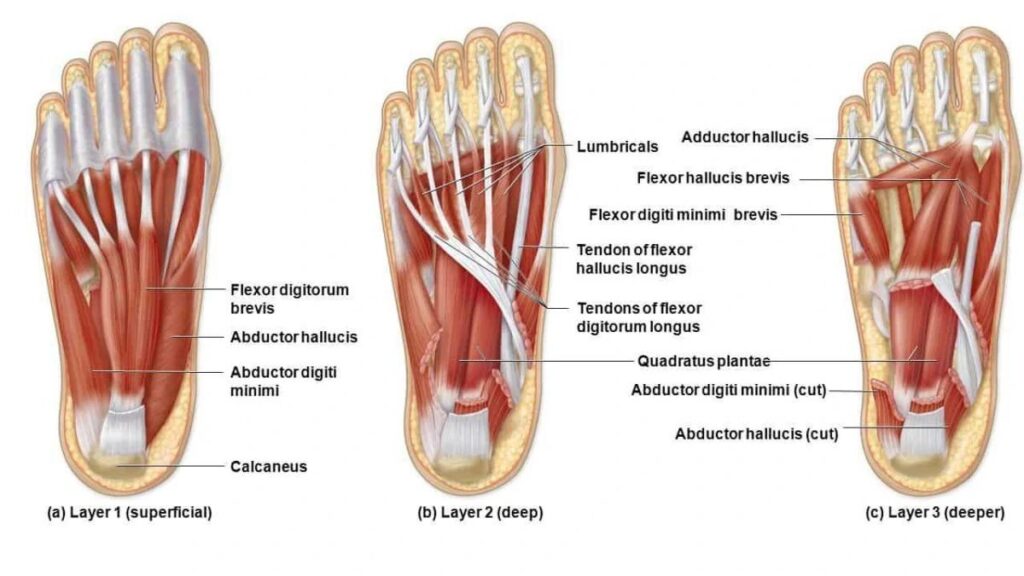
Muscles and Tendons Supporting the Foot
The muscles of the foot are divided into intrinsic and extrinsic groups. Intrinsic muscles are located entirely within the foot. They control small, precise movements, such as adjusting the toes for balance or gripping uneven surfaces.
Extrinsic muscles originate in the lower leg and extend into the foot. These muscles generate the power needed for walking, running, and jumping. Tendons connect these muscles to bones, transmitting force and enabling movement.
One of the most important tendons in the foot is the Achilles tendon, which connects the calf muscles to the heel bone. It allows you to push off the ground when walking or running. Proper function of muscles and tendons is essential for smooth movement, preventing injury, and maintaining overall foot health.
Ligaments and Arches: Nature’s Foot Support System
Ligaments are strong bands of tissue that connect bones and provide stability. The plantar fascia is the longest ligament in the foot, running along the bottom of the foot from the heel to the toes. It supports the arch and absorbs shock during movement.
The foot has three main arches: the medial longitudinal, lateral longitudinal, and transverse arches. These arches act as natural shock absorbers, distributing weight evenly and reducing stress on muscles and joints. When the arches are healthy, they provide a spring-like function that makes walking and running more efficient. Understanding these structures explains why conditions like flat feet or collapsed arches can cause discomfort and affect mobility.
Blood Supply and Nerve Networks of the Foot
Good circulation is vital for healthy feet. The dorsalis pedis artery supplies blood to the top of the foot, while the posterior tibial artery provides blood to the sole. Adequate blood flow ensures that tissues receive oxygen and nutrients, helping prevent injury and promote healing.
The foot also has a complex network of nerves that detect pressure, temperature, and pain. This sensory information helps you maintain balance and react to changes in terrain. Proper circulation and nerve function are critical for overall foot health and mobility.
Common Conditions Linked to Foot Anatomy
Understanding foot anatomy can also help explain why certain problems occur. Structural issues in the bones, joints, or arches can lead to conditions like flat feet, where the arches collapse, causing pain and instability. Plantar fasciitis, a common source of heel pain, occurs when the plantar fascia becomes inflamed, often due to stress or overuse. Bunions are another condition linked to foot structure, forming when the big toe shifts toward the other toes, causing a bony bump.
Awareness of foot anatomy helps prevent these conditions by guiding proper footwear choices, exercise, and injury management. Maintaining foot strength and flexibility supports long-term mobility and reduces the risk of pain or damage.
Conclusion
The foot is an intricate and powerful structure, combining bones, joints, muscles, ligaments, and nerves into a system designed for movement, balance, and support. Understanding foot anatomy helps us appreciate the complexity of this essential body part and highlights the importance of keeping our feet healthy.
Paying attention to foot health—through proper footwear, exercises, and awareness of potential issues—can make a significant difference in daily comfort and long-term mobility. By learning how the foot works, you can take steps to protect it, prevent injuries, and support an active, healthy lifestyle.
FAQs
What is the main function of foot anatomy?
Foot anatomy helps the foot support body weight, maintain balance, absorb shock, and allow smooth movement.
How many bones are in the human foot?
The human foot has 26 bones, including the talus, calcaneus, metatarsals, and phalanges.
What are the arches of the foot, and why are they important?
The foot has three arches—medial, lateral, and transverse—that absorb impact, distribute weight, and maintain stability.
What is the role of the plantar fascia?
The plantar fascia is a ligament along the sole that supports the arches and helps absorb shock during movement.
How can understanding foot anatomy prevent injuries?
Knowing foot anatomy helps you choose proper footwear, perform strengthening exercises, and avoid stress on bones, joints, and ligaments.